
Case Study: Focused Ultrasound Mechanism of Action (MoA) Animation for WVU Rockefeller Neuroscience Institute
How Intervoke built a 3D mechanism of action (MoA) animation showing how focused ultrasound delivers Alzheimer’s drugs for WVU Rockefeller Neuroscience Institute.
Medical Animation Overview
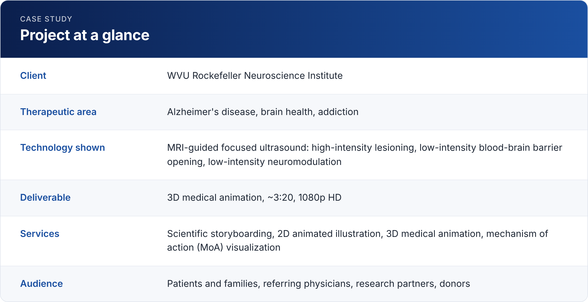
Intervoke produced a 3D mechanism of action (MoA) animation for the WVU Rockefeller Neuroscience Institute (RNI) that explains how MRI-guided focused ultrasound can open the blood-brain barrier to help Alzheimer's drugs reach the brain. RNI ran a first-in-the-world clinical trial pairing focused ultrasound with anti-amyloid antibody therapy, and needed patients, families, clinicians and donors to understand it at a glance.
The animation moves from the MRI suite and transducer helmet down to the blood-brain barrier, microbubbles, antibodies and amyloid plaques. It runs about 3 minutes 20 seconds and is driven by music and on-screen callouts rather than narration.
The challenge
The blood-brain barrier protects the brain, but it also keeps out most medicines. RNI director Dr. Ali Rezai has noted that it limits more than 98% of drugs and therapeutics from reaching the brain from the bloodstream.
RNI's answer is focused ultrasound. Patients wear a helmet with about 1,000 ultrasound transducers, and real-time MRI guides the energy to precise brain regions. Low-intensity ultrasound, together with injected microbubbles, temporarily loosens the barrier so antibody drugs can reach amyloid plaques. In the trial's first three patients, regions where the barrier was opened showed about 32% fewer amyloid plaques after six months than regions left closed.
This mechanism is hard to explain in words. It happens at the cellular scale, inside living tissue, over seconds to hours. RNI needed a healthcare visualization that would:
- Show that the procedure is non-invasive and needs no surgery
- Make the blood-brain barrier understandable to non-scientists
- Show exactly how microbubbles and ultrasound open the barrier, and that it closes again
- Place Alzheimer's drug delivery alongside RNI's other focused ultrasound uses
Our approach: from storyboard to 3D medical animation
In any MoA animation, scientific accuracy comes first, so Intervoke started with hand-drawn 2D storyboards, reviewed with RNI's scientific team, before any 3D work began. Each frame fixed the camera, the scale and the single idea the viewer should take away. That let clinicians check the science early, when changes are cheap.
Three choices shaped the final film:
- One continuous zoom. The camera travels from the MRI suite into the head, into the brain and down to a single capillary. Viewers always know where they are in the body.
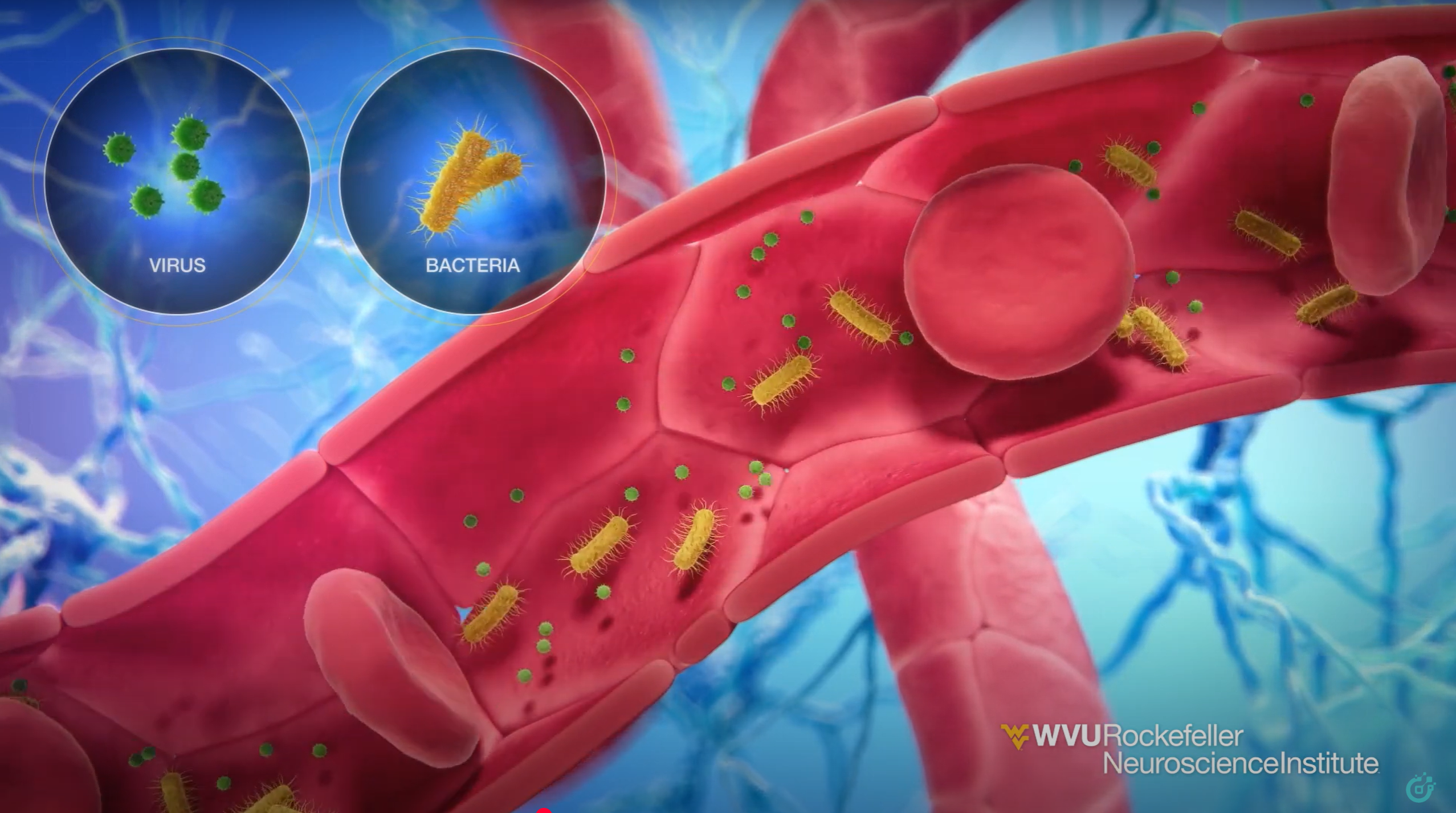
- Clear visual language. Each player has its own color and shape: red blood cells, purple antibodies, blue microbubbles, green pathogens and water molecules. These color keys were set in the storyboard and kept in the 3D renders.
- Music instead of narration. Short on-screen callouts carry the key facts, so the animation works in waiting rooms, at events, on social media and for audiences in any language.
Storyboard to screen: the mechanism of action, scene by scene
The film opens in the MRI suite and introduces three uses of focused ultrasound: high-intensity focused lesioning, low-intensity blood-brain barrier opening for targeted drug delivery, and low-intensity neuromodulation. A patient slides into the scanner, and a callout reads "A non-invasive procedure requiring no surgery." The storyboard frames below cover the heart of the film, the Alzheimer's drug delivery sequence, in the order they appear.
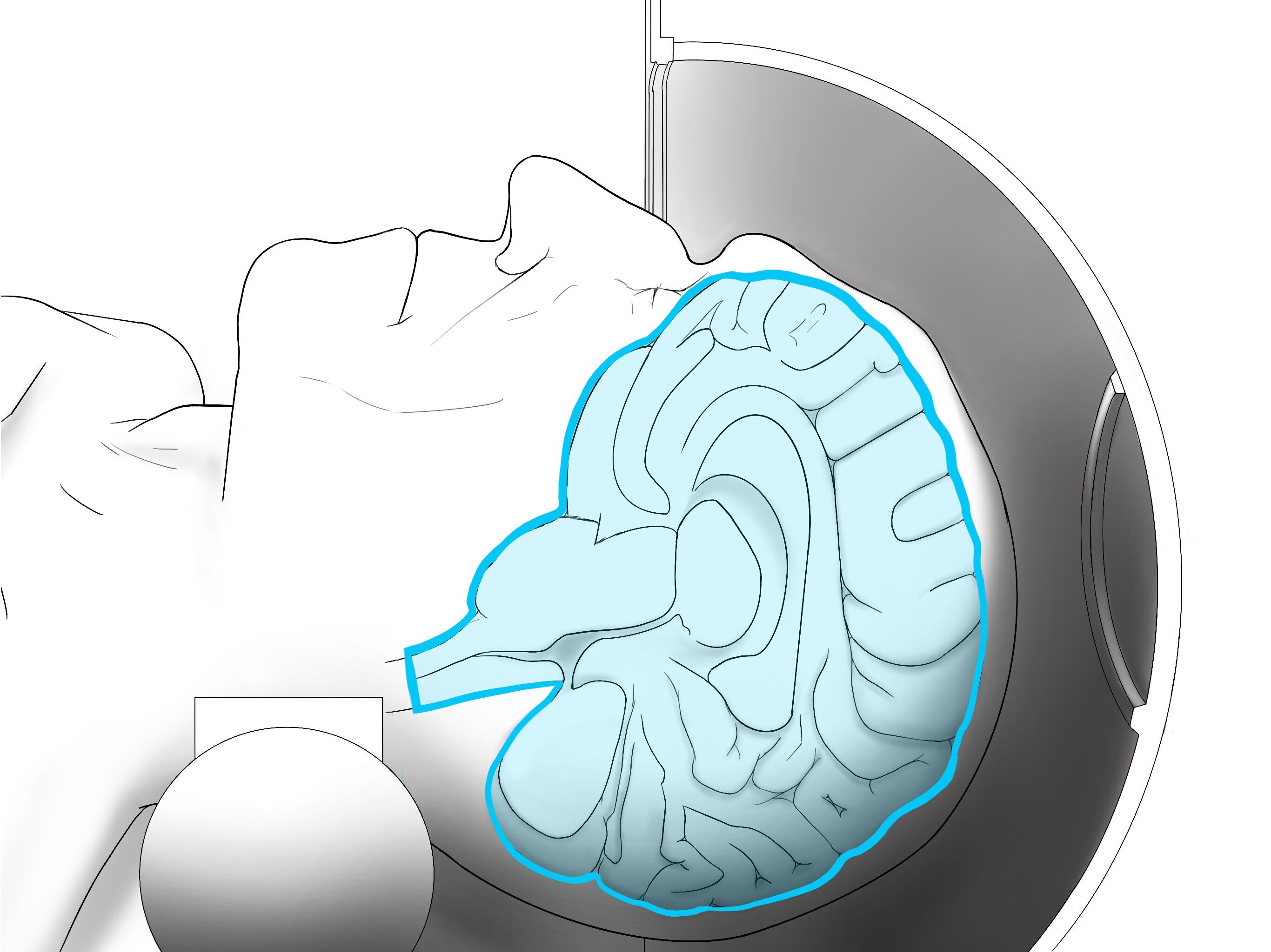
1. The brain inside the helmet (about 0:30)
A cutaway reveals the patient's brain inside the ultrasound helmet. The storyboard outlined the whole brain in a single highlight color so viewers read it instantly against the line-art patient and scanner.
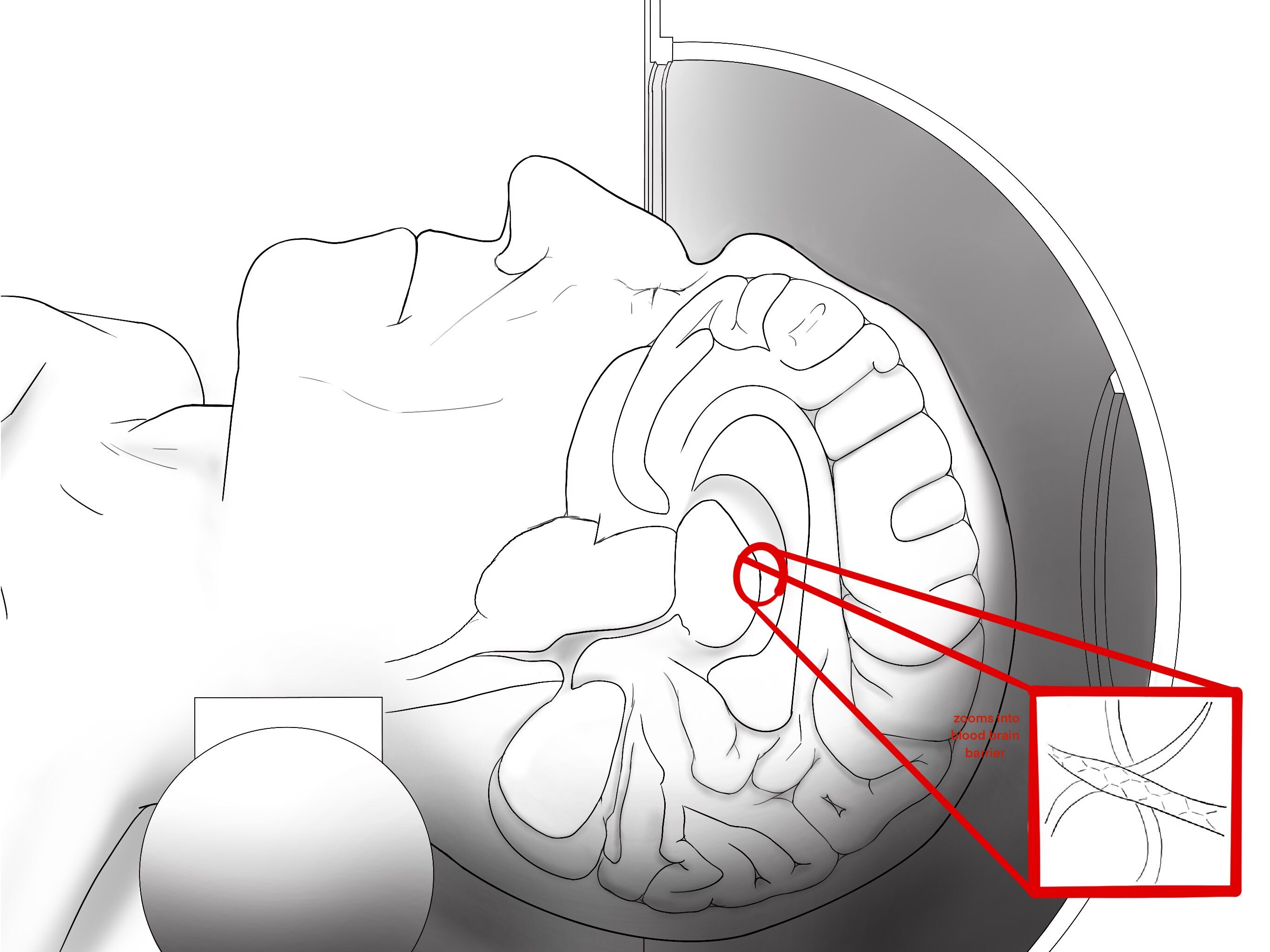
2. Zooming into the blood-brain barrier (about 1:00)
A magnifier marks one deep target, and the camera dives in to a single capillary. The storyboard note simply reads "zooms into blood brain barrier," and the final cut follows it as one unbroken move.
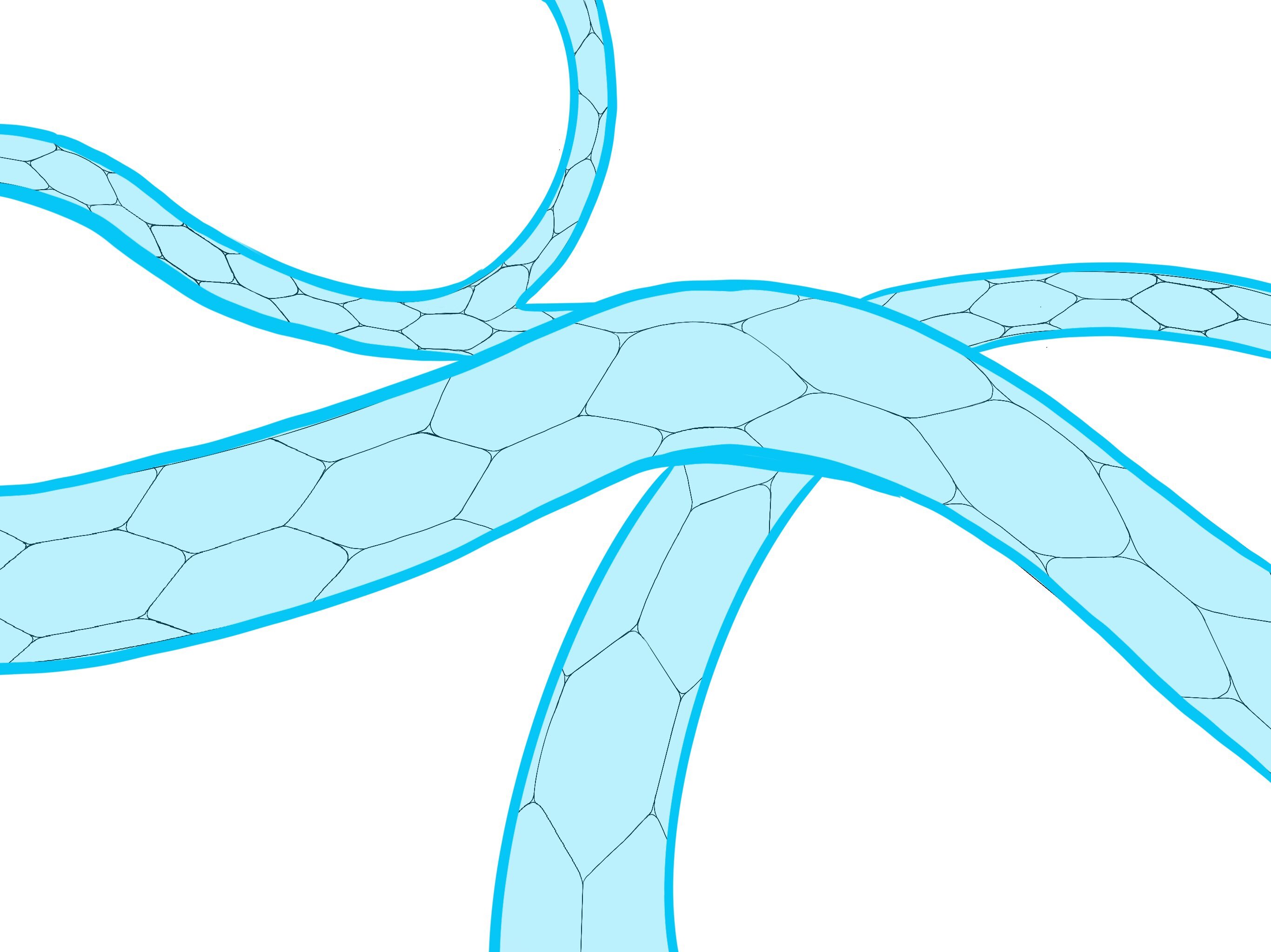
3. The brain's capillary network
The first look at the vessels shows their tightly fitted endothelial cells, drawn as a honeycomb of tiles. That pattern sets up the "tight junctions" the rest of the story depends on.
4. A barrier that protects (about 1:05)
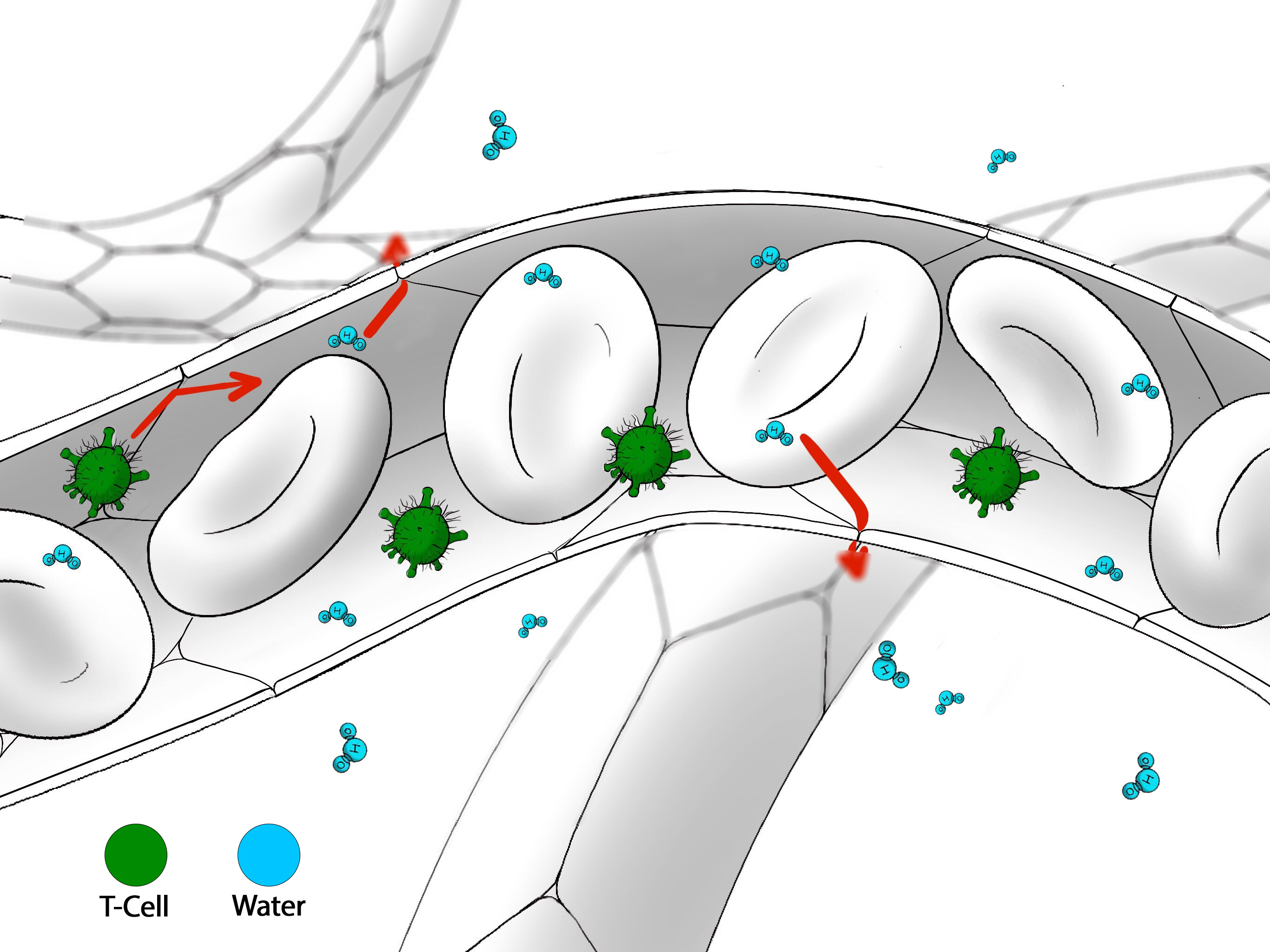
Inside the vessel, red blood cells flow past viruses and water molecules. Water slips through the vessel wall into the brain, while viruses bounce off. In the 3D animation, a "Virus" callout labels the pathogens.
5. A barrier that blocks treatment (about 1:20)
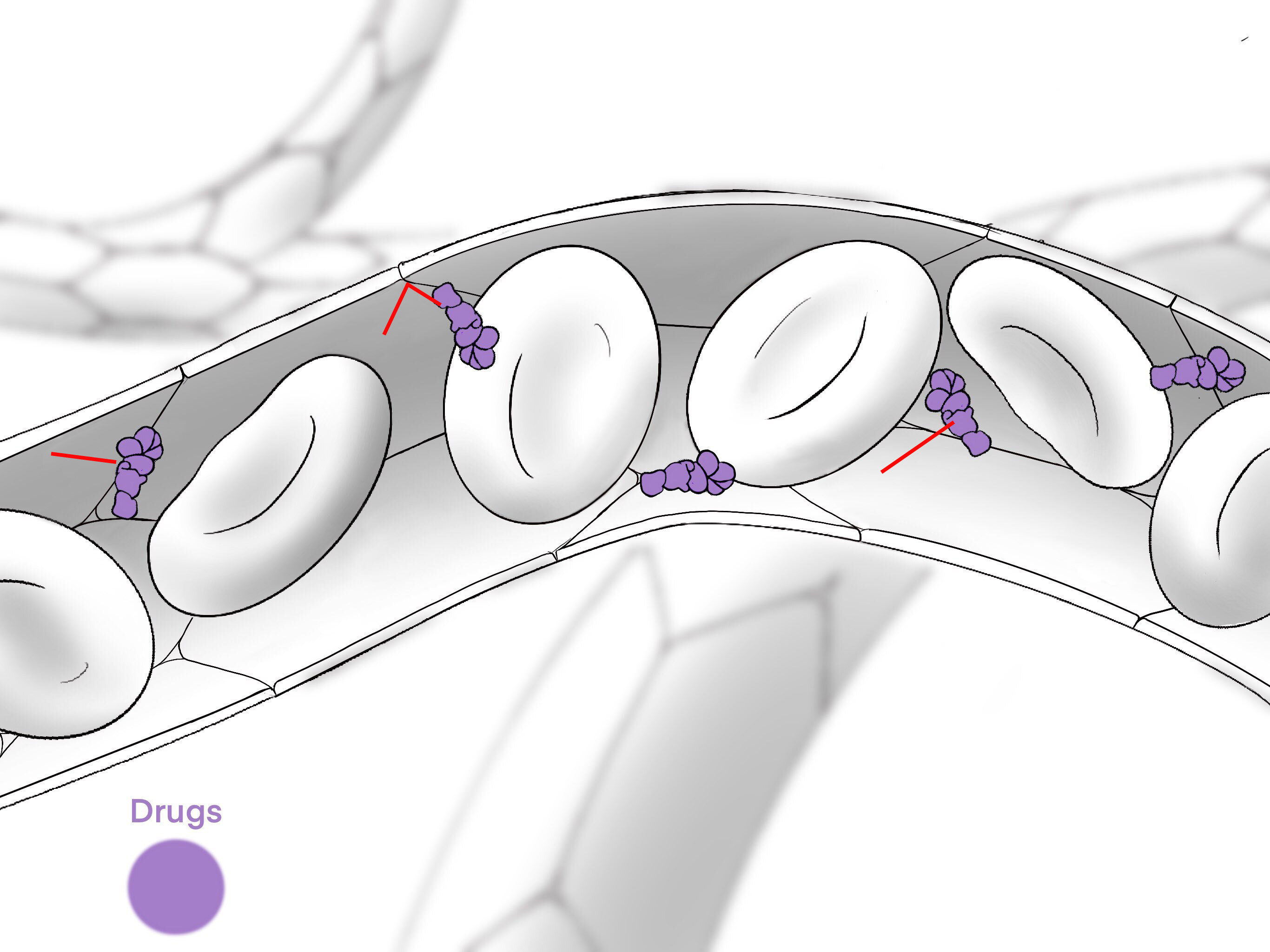
The same wall that stops viruses also stops antibody drugs. Purple antibodies, labeled "Antibody" in the final film, gather along the vessel wall but cannot cross it.
6. MRI-guided focused ultrasound (about 1:35)
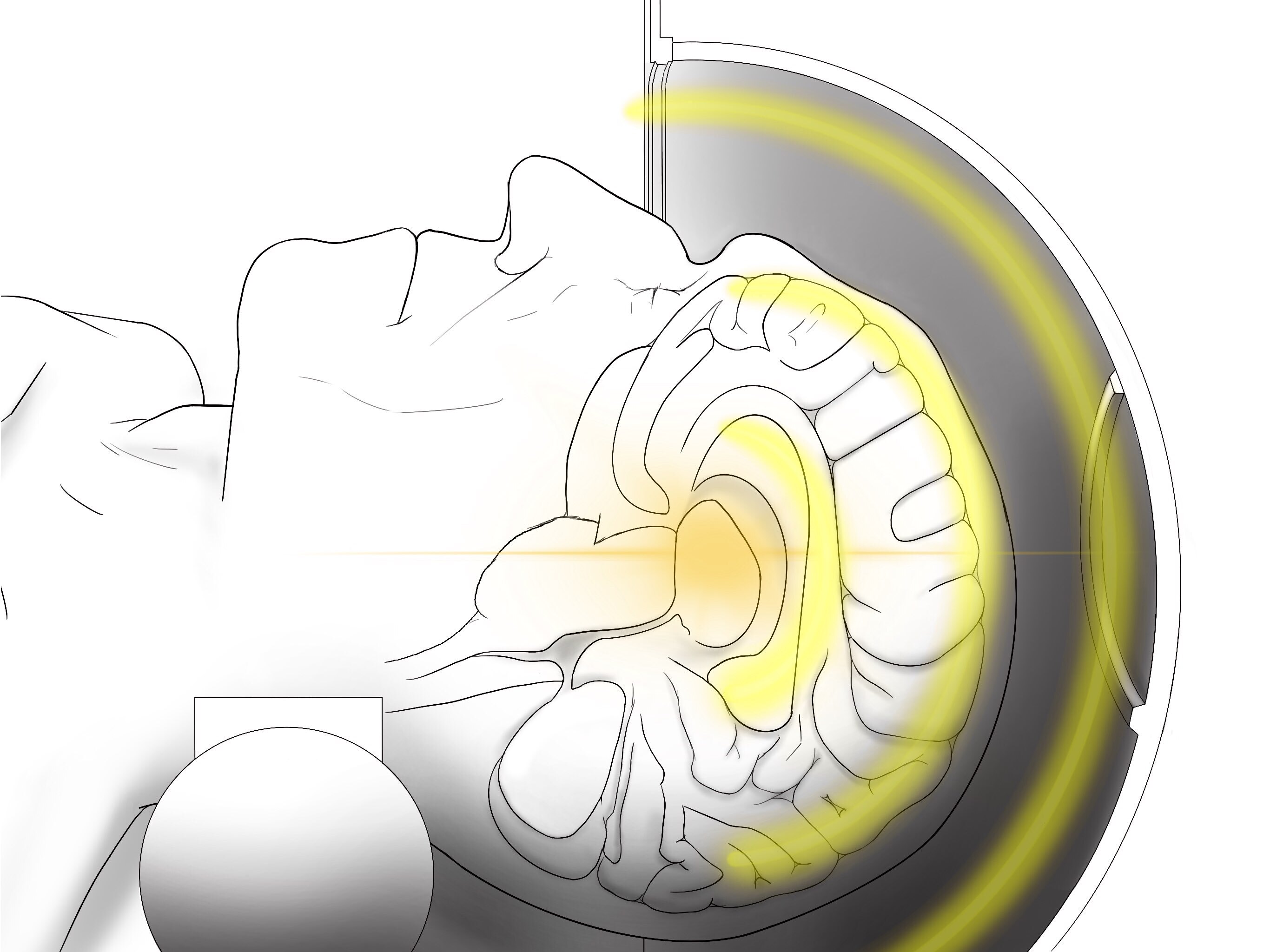
The camera pulls back to the helmet, and ultrasound waves converge on a single point in the brain. Callouts state that the helmet uses 1,000 ultrasound transducers and MRI-guided brain targeting.
7. Microbubbles open the barrier (about 1:50)
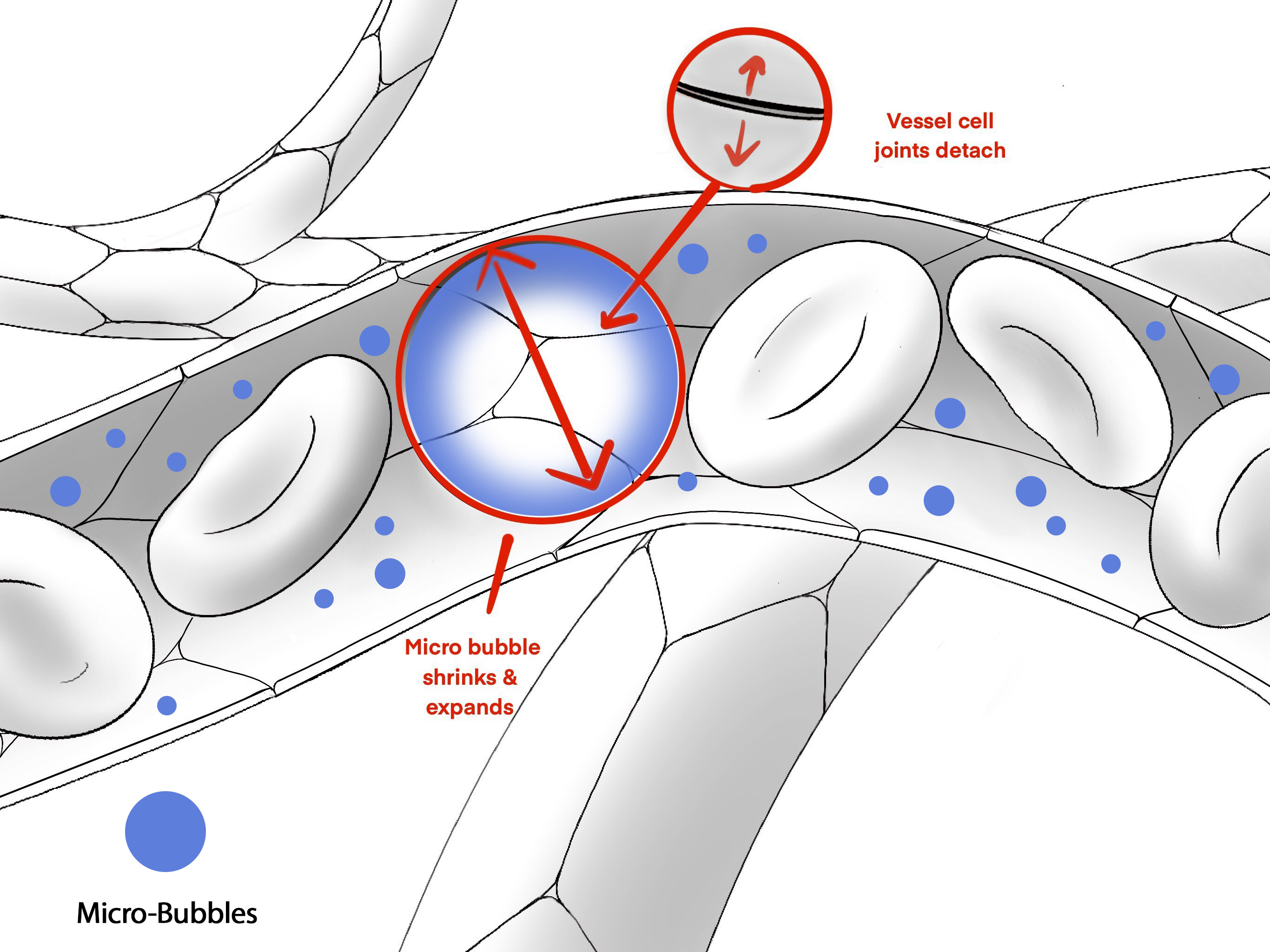
Microbubbles in the bloodstream shrink and expand as the ultrasound hits them. This gentle push separates the joints between vessel cells. The storyboard labels both steps, "Micro bubble shrinks & expands" and "Vessel cell joints detach," and the 3D render shows a bubble swelling until a seam opens in the wall.
8. The drug reaches its target (about 2:05)
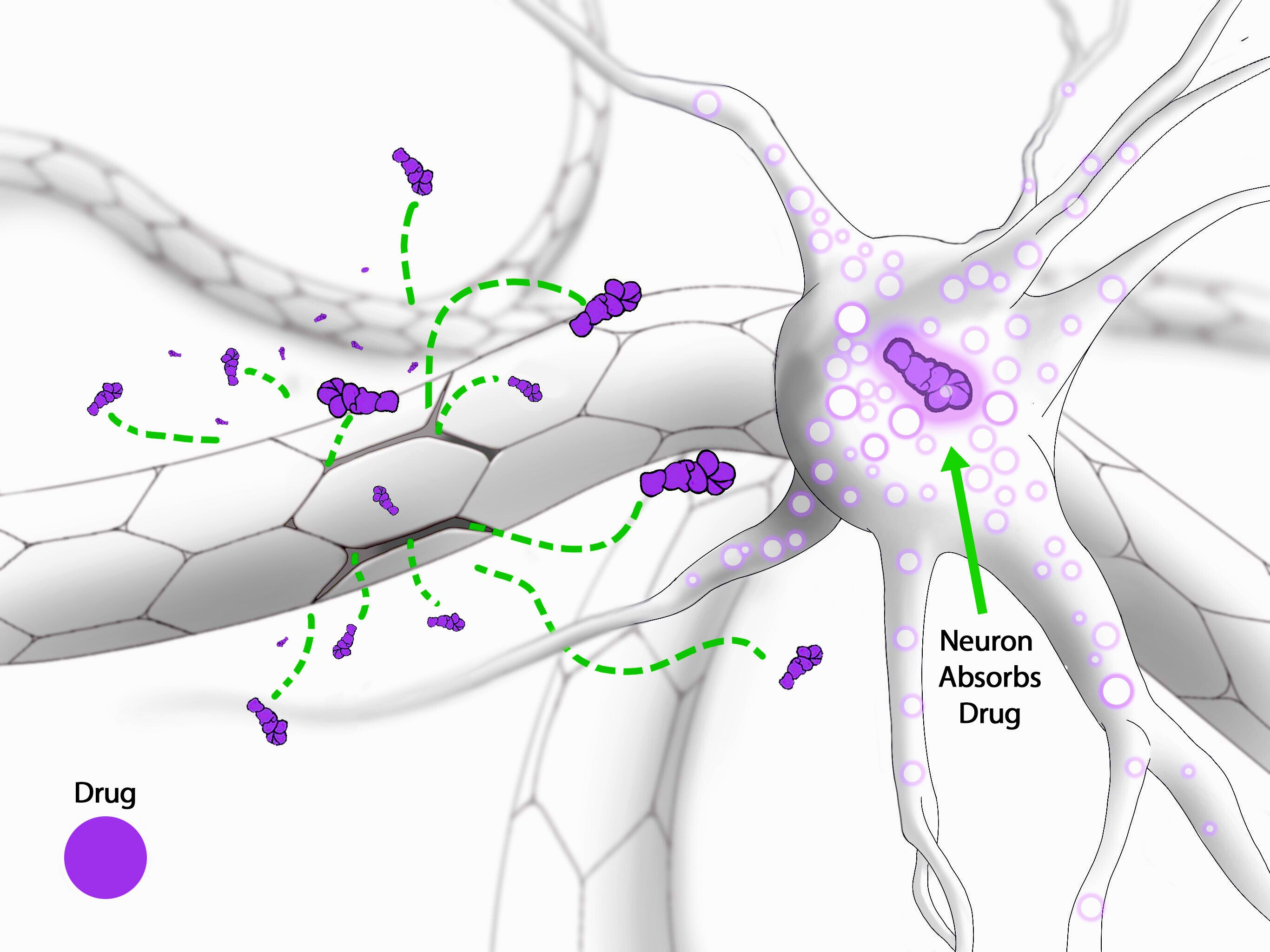
Antibodies flow through the open seams toward amyloid "Plaque" deposits and the neurons around them, and are taken up in the brain. Two clock overlays, 8 hr and 38 hr, then show the vessel cells sealing back together, making clear that the opening is temporary.
Beyond Alzheimer's: lesioning and neuromodulation
The animation also sets RNI's Alzheimer's work within its wider focused ultrasound program, so one asset can serve several service lines.
- High-intensity focused lesioning. A target ring locks onto one point deep in the brain, then a small dark spot forms where the beam converges, showing a precise, incision-free lesion.
- Low-intensity neuromodulation. Icons for drugs, alcohol, gambling and other compulsions radiate from a reward center that glows red, signaling addiction-related activity. Ultrasound pulses calm the overactive neurons, which shift from pink to blue. Icons of healthy activities, such as exercise, family and connection, then take their place.
The film closes on the patient resting in the scanner and the WVU Rockefeller Neuroscience Institute logo.
The result
RNI now has one 3D medical animation that explains a complex mechanism of action (MoA) in about three minutes, without a single spoken word. It gives the institute:
- A patient education tool that shows people considering a clinical trial what will happen, and reassures them that the procedure needs no surgery and the barrier closes again
- A physician and partner explainer that makes MRI-guided focused ultrasound and targeted drug delivery easy to grasp in a meeting or a talk
- Reusable assets. The 3D models of the helmet, brain, capillaries, microbubbles and antibodies can be reus
Bring your science to life
Intervoke creates 2D and 3D medical animation, MoA and MoD animation, medical device animation and interactive healthcare education applications for the pharmaceutical, biotech, medical device and academic medicine sectors. If your team has a breakthrough that is hard to put into words, book a discovery call to talk about your next project.
Sources
- WVU RNI expands groundbreaking Alzheimer's research in latest trial, My Buckhannon
- How Focused Ultrasound is Changing Alzheimer's Treatment, BrightFocus Foundation webinar with Dr. Ali Rezai, May 15, 2025
- Focused ultrasound plus plaque-reducing drugs could slow Alzheimer's progression, Physics World
- Intervoke
Let’s Get Started
Interested in learning more about how Intervoke can bring your science to life?
Contact us or book a Discovery Call to get started.


