
Hyalex Freestyle Knee Implant: A 3D Mechanism of Action Animation for Cartilage Repair
How Intervoke used 3D mechanism of action animation to show how the HYALEX knee implant restores damaged cartilage, from disease to surgery.
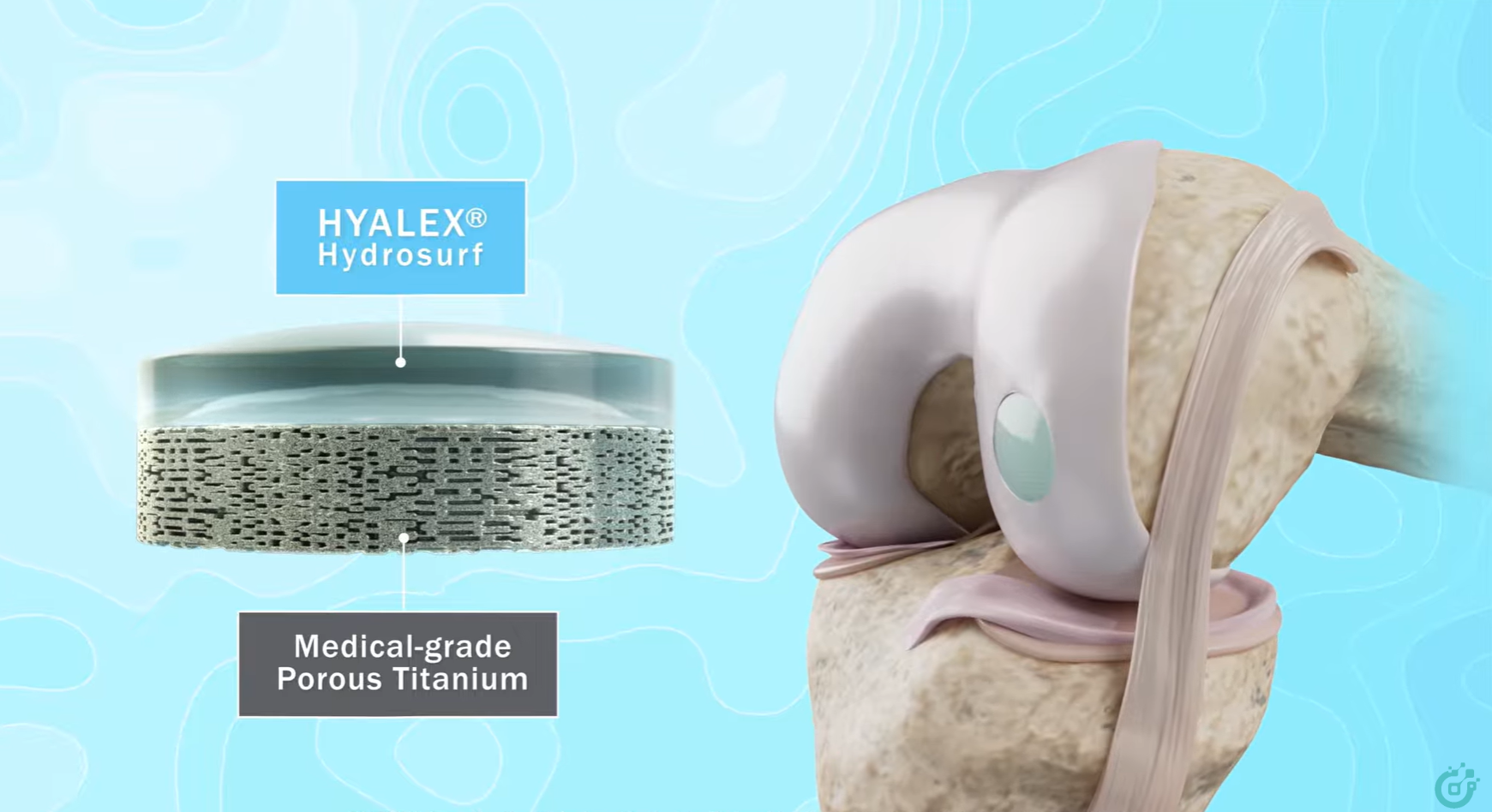
At a glance
Intervoke produced a 70–80 second 3D mechanism of action (MoA) animation for Hyalex Orthopaedics. It shows how the HYALEX Freestyle Knee Implant restores damaged knee cartilage in a single surgery: what the implant is made of, how it bonds with bone, and how surgeons place it step by step.
The challenge
Hyalex Orthopaedics is a medical device company based in Lexington, Massachusetts. It is building joint-preservation technology for patients with knee cartilage damage and early osteoarthritis, a group with few treatment options today. The Freestyle Knee Implant uses Hyalex's patented HYDROSURF material, which is designed to match the structure and function of natural hyaline cartilage.
The product's key benefits happen out of sight. A surgeon can't watch a synthetic cartilage surface move with the joint, or bone grow into a titanium base. Hyalex needed one short video that could:
- Explain why damaged hyaline cartilage needs repair: it can't heal itself.
- Show how the implant works: a biomimetic cartilage surface on a porous titanium base that treats cartilage and the bone beneath it.
- Walk surgeons through the procedure: sizing, guide pin, drilling, trialing, loading and seating the implant.
- Work on a booth screen, in an email and in a social feed, all from a single asset.
Our approach
We followed our usual three-phase process for medical device animation. Each phase had a sign-off, so the science was settled before anything was rendered.
Phase 1: Script and storyboard. Our scientific writers wrote a script of about 80 seconds, timed at 150–160 words per minute so the voiceover would be clear without feeling rushed. Hyalex's biomedical engineering team reviewed it with tracked changes and references. We then built a 10-scene PowerPoint storyboard with narration, on-screen action and look development for every shot.
Phase 2: Voiceover, 3D modeling and animation. Hyalex supplied CAD files for the implant and the full Freestyle instrument set: pin guide, guide pin, drill, drill guide, trial, inserter sleeve, tamp, mallet and sterile packaging. We optimized them for animation so every instrument looks exactly like the real one. We added existing Intervoke 3D anatomy IP to save time and cost. Rough animatics went to Hyalex on a password-protected Vimeo page, where the team left time-stamped comments on camera timing, labels and transitions.
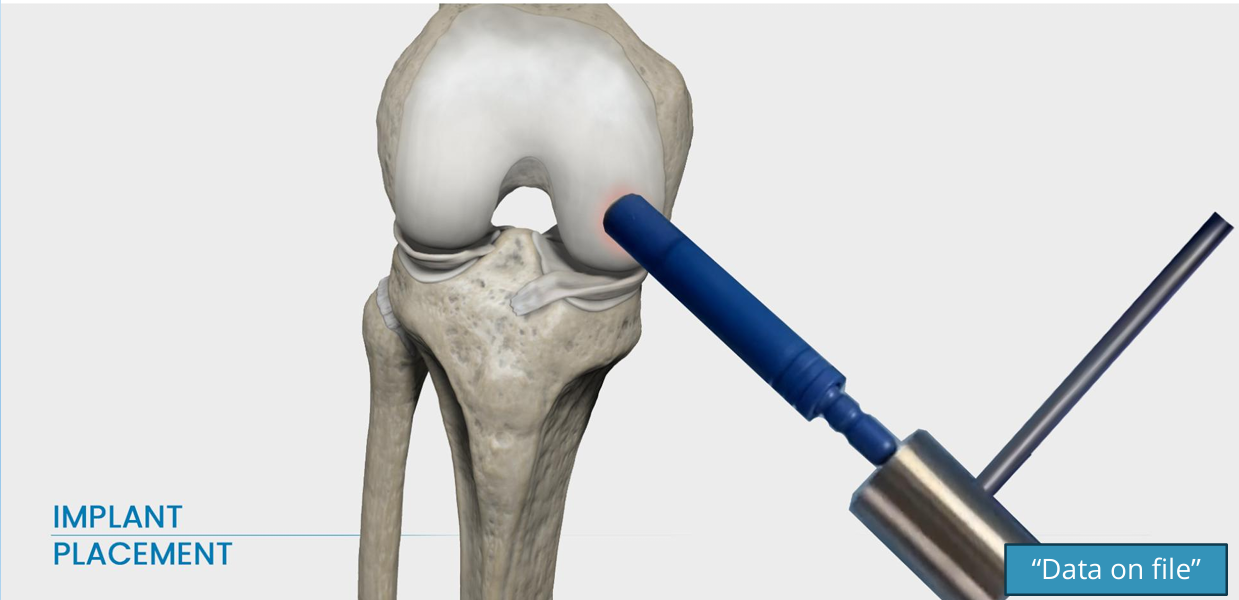
Phase 3: Rendering and post-production. We rendered final layers on our GFX render farm, then composited them with visual effects, music and sound design. Branded labels and the required "Data on file" note were added to the frames that make product claims.
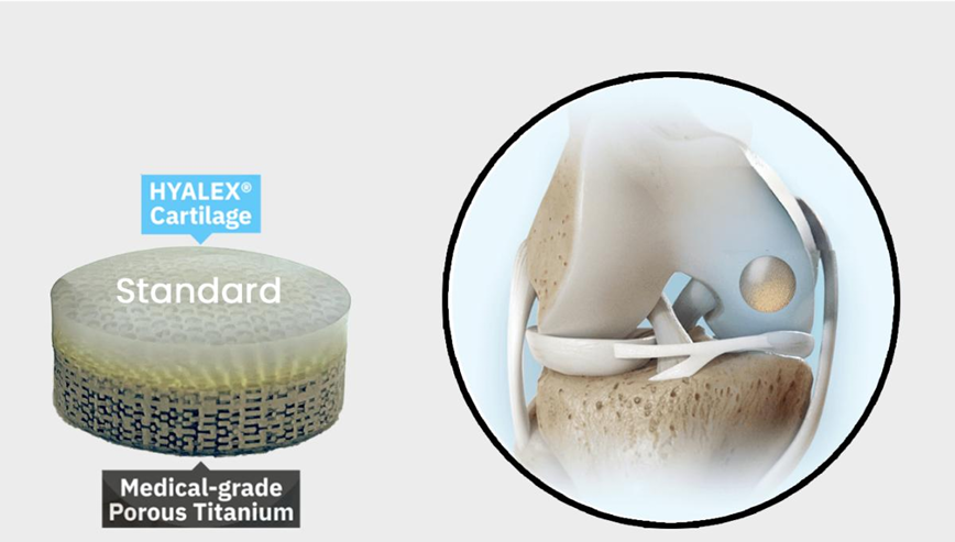
Telling the mechanism of action story
The animation has three acts. Together they answer the questions a surgeon will ask: what's wrong, how the implant fixes it, and how to place it.
1. Mechanism of disease: cartilage that can't heal
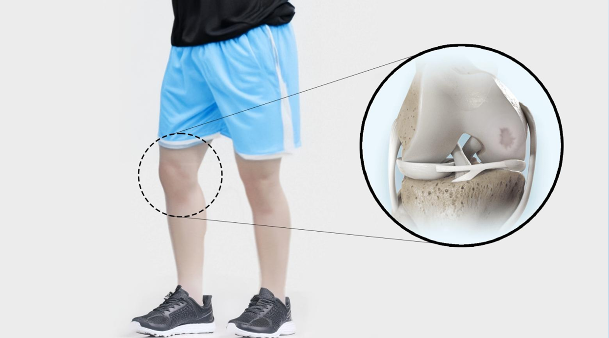
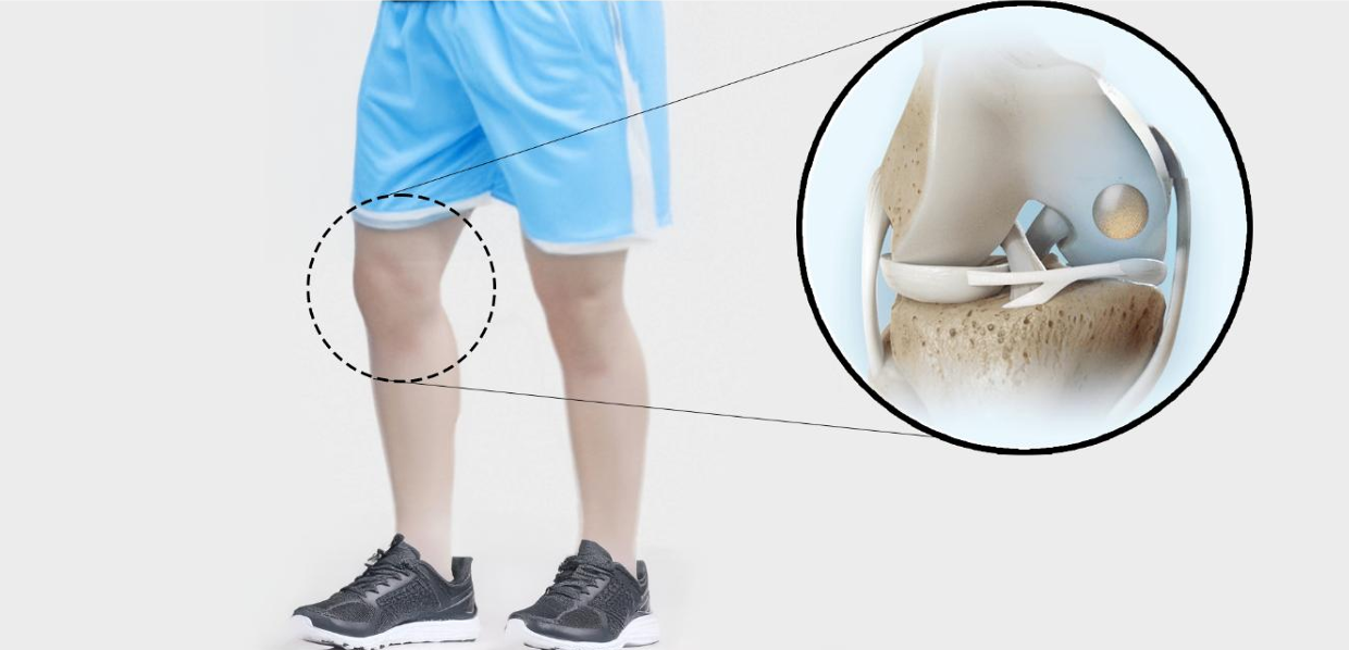
The film opens on a runner's knee flexing smoothly. A zoomed callout shows healthy hyaline cartilage at work. As he slows down, the joint ages over time and an isolated cartilage lesion forms on the femoral condyle. The narration makes the clinical point: once damaged, hyaline cartilage can't heal itself, and lesions cause pain and inflammation.
2. Mechanism of action: a biomimetic implant with a titanium base
Next, the HYALEX implant rotates on its own so viewers can see both layers: the HYDROSURF biomimetic cartilage surface and the medical-grade porous titanium base. On-screen text and a millimeter scale show that it comes in 16 sizes for a wide range of lesions. The key MoA moment comes after placement. A sagittal cross-section shows the implant surface lining up with the native cartilage while bone grows into the titanium pores. This shows how the press-fit gives stability right away and bone ingrowth over time.
3. Surgical technique: six steps, one surgery
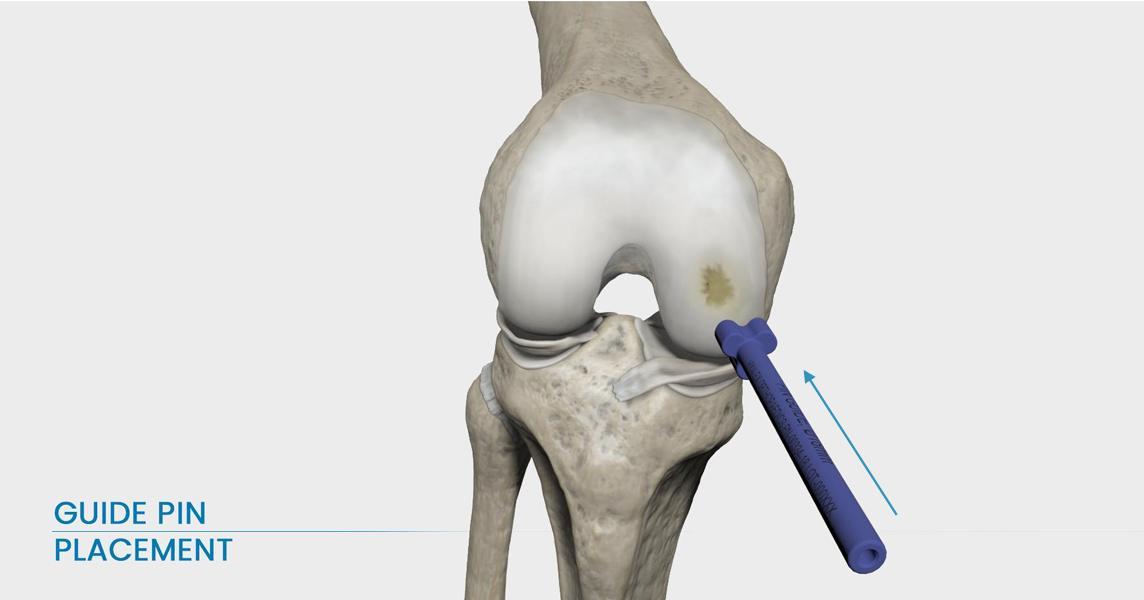
The procedure section moves quickly, at about five seconds per step. The skin fades away and each CAD-accurate instrument moves into place:
- Access and sizing: the pin guide sizes the lesion and sets the implant size.
- Guide pin placement: a pin is drilled into the center of the lesion.
- Site preparation: the drill removes damaged cartilage and bone over the pin.
- Depth verification: the trial confirms a flush fit.
- Implant loading: the implant goes from its packaging into the inserter sleeve.
- Implant placement: the tamp and mallet seat the implant with three taps.
The film ends where it started, with the runner moving freely in slow motion. A final zoom shows the implant working inside the repaired joint.
The result
Hyalex received one 1080p animation that takes viewers from the disease, to the device's mechanism of action, to the full surgical technique in under 90 seconds. Because it was built from Hyalex's own CAD data, every instrument matches the real kit, so the film works both as a surgeon education tool and as a marketing asset. The finished video now supports Hyalex's website, social channels, email outreach and trade show booth. The 3D models and source files can be reused for future sales aids, presentations and interactive projects.
Frequently asked questions
What is a mechanism of action animation for a medical device?
A mechanism of action (MoA) animation shows how a device produces its effect inside the body: what it's made of, how it works with tissue, and why that matters clinically. For the HYALEX implant, that means showing how a biomimetic cartilage surface restores the joint and how a porous titanium base supports bone ingrowth.
How does the HYALEX Freestyle Knee Implant work?
The implant has a HYDROSURF biomimetic surface designed to match natural hyaline cartilage, on top of a porous titanium base. Surgeons press-fit it into a drilled site on the femoral condyle. This treats both the cartilage lesion and any damaged bone beneath it in a single surgery, and bone grows into the titanium over time.
Why use 3D animation to explain an orthopedic implant?
The key events, such as cartilage wear, implant fit and bone ingrowth, happen where no camera can go. 3D animation can fade away skin, cut cross-sections and slow down time, so surgeons and patients can see exactly how a device works.
How long does it take to produce a medical device animation?
This project followed Intervoke's three-phase process: script and storyboard, then 3D modeling and animation, then rendering and post-production. It was scheduled over about eight weeks from kickoff to final delivery. Timelines depend on length, detail and how many review rounds are needed.
Can CAD files be used in medical animation?
Yes. Hyalex supplied CAD models of the implant and nine instruments. Intervoke optimized them for animation, so every tool on screen matches the real surgical kit.
Bring your device's mechanism of action to life
Whether you're launching an orthopedic implant, a surgical instrument or a new therapy, Intervoke turns complex science into clear, accurate 3D medical animation. Schedule a Discovery Call today.
Let’s Get Started
Interested in learning more about how Intervoke can bring your science to life?
Contact us or book a Discovery Call to get started.


